|

|

|

| Robert Weinreb (chair)
| David Friedman (co-chair)
| Paul Foster (co-chair)
| Aung Tin (co-chair)
 Reports for this consensus were prepared and discussed using the same
internet based e-Room system as used with the previous two reports. The
consensus book on Angle Closure Glaucoma is published by
Kugler Publications
(see below for details). Reports for this consensus were prepared and discussed using the same
internet based e-Room system as used with the previous two reports. The
consensus book on Angle Closure Glaucoma is published by
Kugler Publications
(see below for details).
The Consensus Faculty consisted of the leading authorities in Angle
Closure with representatives from six continents. These 110 experts
dedicated their knowledge, time, and insight to the preparation of the
reports between January 1 and May 1, 2006. Prior to the meeting, each of
the WGA member Glaucoma Societies was sent a draft of the consensus
report for comment. Each member Society was also invited to send a
representative to attend the consensus meeting. The report then was
discussed extensively during the Consensus Meeting. Reports and
Consensus Statements were revised following these discussions.
Epidemiology, classification and mechanism
Classification
- The proposed classification scheme can be used not only to classify
the natural history of angle closure, but also to determine prognosis
and describe an individual's need for treatment at different stages of
natural history of the disease.
- Additional clinical sophistication can be gained describing sequelae
of angle closure affecting the cornea, trabecular meshwork, iris, lens
optic disc and retina. Specifically, the extent of peripheral anterior
synechiae (PAS), level of presenting IOP (in asymptomatic cases) and presence of glaucomatous optic
neuropathy.
- Ascertaining the mechanism of angle closure (pupillary block, plateau,
lens-related, retro-lenticular) is essential for management, and it
should be used in conjunction with a classification of the stage of the
disease.
Comment: Further refinement of these systems (such as the inclusion of
symptoms as a defining feature of angle closure) should be made on the
basis of peer-reviewed evidence.
Gonioscopy
- Gonioscopy is indispensable to the diagnosis and management of all
forms of glaucoma and is an integral part of the eye examination.
- An essential component of gonioscopy is the determination that
iridotrabecular contact is either present or absent. If present, the
contact should be judged to be appositional or synechial (permanent).
Comment: The terms 'iridotrabecular contact (and number of degrees) and
'primary angle closure suspect' should be substituted for 'occludable',
as more accurate.
Comment: The determination of synechial contact may require indentation
of the cornea during gonioscopy, in which case a goniolens with a
diameter smaller than the corneal diameter is preferred.
- Access to a magnifying, Goldmann-style lens enhances the ability to
identify important anatomical landmarks, and signs of
pathology. Although the accuracy of indentation with this lens has not
been validated, its use does complement that of a goniolens with a
diameter smaller than the corneal diameter. The ideal standard is access
to both types of lens.
- Anterior segment imaging devices may augment the evaluation of the
anterior chamber angle, but their place in clinical practice still needs
to be determined.
- It is desirable to record gonioscopic findings in clear text.
Describing the anatomical structures seen, the angle width, the iris
contour and the amount of pigmentation in the angle are all desirable.
Issues requiring further attention
- Develop a specific definition of PAS.
- Re-consider of the definition of a primary angle closure suspect to
include those with any irido trabecular contact (ITC) or perhaps 180� of
ITC, as the current definition (which requires 270� of ITC) excludes
around 50% of cases with primary angle closure causing PAS.
- Inclusion of disc size when seeking structural changes consistent with
glaucoma in the diagnostic algorithm for future epidemiological studies.
Mechanisms
- Identification of the underlying cause of angle closure is essential
to accurate diagnosis and treatment.
Comment: Angle closure can be caused by one or a combination of
abnormalities in the relative or absolute sizes or positions of anterior
segment structures or abnormal forces in the posterior segment that may
alter the anatomy of the anterior segment. Angle closure may be
understood by regarding it as resulting from blockage of the trabecular
meshwork caused by forces acting at four successive anatomic levels: the
iris (pupillary block), the ciliary body (plateau iris), the lens
(phacomorphic glaucoma), and vectors posterior to the lens (malignant
glaucoma).
- Although the amount of pupillary block may vary among eyes with angle
closure, all eyes with angle closure require treatment with iridotomy.
Management of acute angle closure crisis
- Laser peripheral iridotomy (LPI) should be performed as soon as
feasible in the affected eye(s), and should also be performed as soon as
possible in the contralateral eye.
- Medical management is the recommended first step in treating acute
angle closure, but the results of studies comparing this to immediate
laser surgery are not yet available.
- Laser iridoplasty can be effective at breaking acute attacks
and should be considered if an attack cannot be broken by other means.
- Paracentesis should be reserved for cases where other approaches have
failed.
- Primary cataract extraction may be a treatment option, but data
supporting its use are limited.
Surgical management of primary angle closure glaucoma
- Laser peripheral iridotomy (LPI) is recommended as the primary
procedure in eyes with primary angle closure glaucoma (PACG.
Comment: LPI can be performed easily on an outpatient basis and patients
can then be monitored for response to treatment. This will allow time to
undertake elective surgery in those with uncontrolled IOP, those with
advanced disease or with co-existing cataract. LPI also serves as
prophylaxis against acute angle closure.
- There is lack of evidence for recommending primary incisional surgery
(without LPI) in eyes with PACG.
- Trabeculectomy may be performed to lower IOP in eyes with chronic
PAC(G) insufficiently responsive to laser or medical therapy.
- There is insufficient evidence for deciding which cases with PACG
should undergo cataract surgery alone (without trabeculectomy).
Comment: Cataract surgery alone may be considered in eyes with mild
degree of angle closure (less then 180� degrees of PAS), mild optic
nerve/visual field damage or those that are not on maximal tolerated
medical therapy.
- There is lack of evidence for recommending lens extraction alone in
eyes with more advanced PACG.
Comment: Published studies to date have been non-randomized with small
sample sizes and short follow-up.
- Combined cataract and glaucoma surgery in certain eyes may be useful
to control IOP and restore vision.
Comment: There is limited published evidence about the effectiveness of
combined cataract extraction and trabeculectomy in eyes with PACG. There
is a need for studies comparing this form of surgery with separately
staged cataract extraction and trabeculectomy
- There is limited evidence about the effectiveness of
goniosynechialysis in the management of PACG.
Laser and medical treatment of primary angle closure glaucoma
- Medical treatment should not be used as a substitute for Laser
peripheral iridotomy (LPI) or surgical iridectomy in patients with PAC
or PACG.
- Prostaglandin analogues appear to be the most effective medical agent
in lowering IOP following LPI, regardless of the extent of synechial
closure.
Detection of primary angle closure and angle closure glaucoma
- Angle closure case detection or opportunistic screening should be
performed in all persons forty years of age and older undergoing an eye
examination.
- Given the low specificity of the flashlight test, it is not
recommended for use in population-based screening or in the clinic.
- A shallow anterior chamber is strongly associated with angle closure.
The use of anterior chamber depth (ACD) defined here again for
population-based screening is as yet unproven.
- Many clinicians currently perform iridotomy as prophylaxis in the
presence of any visible irido trabecular contact (ITC).
Comment: Published evidence is lacking to justify this practice since it
is unknown whether LPI is effective at preventing AAC, PAC, and PACG
from developing in individuals with gonioscopically detected ITC.
Comment: Research is needed to determine racial/ethnic variations in
response to iridotomy.
Comment: Evidence is needed to evaluate the meaning of a shallow limbal
anterior chamber depth (LACD) in the presence of an 'open' angle on
gonioscopy.
- There is currently no evidence in the literature supporting the
standard use of provocative tests for angle closure. A negative
provocative test does not exclude angle closure.
WGA Consensus
Meetings Publications
|
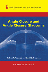
Consensus Series 3
Angle Closure and Angle Closure Glaucoma, edited by By:
R.N. Weinreb & D.S. Friedman
|

|
Publication year: 2006. xiv and 98 pages with 59
figures of which 3 in color, 1 table. Hardbound.
ISBN 10: 90-6299-210-2 ISBN 13:
978-90-6299-210-2
€ 45.00 / US $ 56.00
Available at your local bookstore and directly from the
publisher.
Click here for details
On IGR web: 3rd
WGA Consensus Meeting statements: Angle Closure and
Angle Closure Glaucoma
|
|
Consensus Series 2
Glaucoma Surgery: Open Angle Glaucoma, edited by Robert N.
Weinreb and Jonathan G. Crowston
|

|
Publication year: 2005. xiv and 140 pages with 9 tables and 2
figures, of which 1 in full color Hardbound.
Kugler Publications, The Hague, The Netherlands.
ISBN 10: 90-6299-203-X ISBN 13: 978-90-6299-203-4
€ 50.00 / US $ 60.00
Available at your local bookstore and directly from the
publisher.
Click here for details
On IGR web: 2nd
WGA Consensus Meeting statements: Glaucoma Surgery:
Open Angle Glaucoma
|
|
Consensus Series 1
Glaucoma Diagnosis. Structure and Function, edited by
Robert.N. Weinreb & Erik. L. Greve
|

| Publication year: 2004. viii and 152 pages with 17 figures,
of which 12 in full color, and one table. Hardbound.
Kugler Publications, The Hague, The Netherlands.
ISBN 10: 90-6299-200-5 ISBN 13: 978-90-6299-200-3
€ 50.00 / US $ 60.00
Available at your local bookstore and directly from the
publisher.
Click here for details
Meeting pictures
|
Issue 8-2
Change Issue
advertisement

| | |

